The Functional Organization of The Lateral Habenula and its Relationship to MDD (Depression)
Project Overview
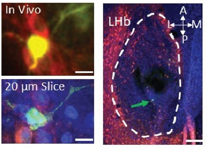
By imaging lateral habenula (LHb) neurons in awake, behaving mice, we discovered that a specific subset of these neurons conveys temporal-difference (TD) error-like signals. However, it remains unclear how this functional heterogeneity maps onto the known molecular and projection diversity of the LHb. To bridge this gap, we developed a highly precise pipeline that combines in vivo imaging with ex vivo histology (Fig. 4). This allows us to track live functional activity and then definitively map those precise signals onto the distinct molecular identities and projection targets of the neurons we observed.
In this project, our lab seeks to dissect this complex functional architecture using state-of-the-art methodology. We aim to determine how distinct populations of LHb neurons are organized to compute these specific aversive error signals, and to uncover how the transcriptional profiles of these exact cell types are fundamentally altered in the context of Major Depressive Disorder (MDD).
Methodology
- Two-Photon Imaging & Photoactivation: Combining two-photon calcium imaging (using a red indicator) in awake, behaving mice with two-photon activation of photoactivatable GFP. This allows us to perfectly register live in vivo functional images with post-mortem histological slices (Fig. 4).
- Multiplexed FISH: Performing multiplexed fluorescence in situ hybridization using 12 specific markers derived from scRNA-seq studies to definitively establish the molecular identities of the photoactivated neurons.
- Targeted Projection Mapping: Categorizing the molecular identities of LHb neurons that project specifically to dopaminergic and GABAergic neurons, allowing us to predict downstream, cell-type-specific targets based purely on in vivo imaging profiles.
- Human snRNA-Sequencing: Sequencing single nuclei from the postmortem human habenula, utilizing highly valuable tissue samples obtained specifically from unmedicated MDD patients.
Current Objectives
We are currently processing our murine data to establish a reliable model that predicts cell-type-specific downstream targets based on the molecular identity of the LHb neurons we image in vivo. In parallel, we plan to combine these functional mouse maps with our snRNA-sequencing data from human patients to definitively determine whether—and exactly how—these specific TD error-encoding habenula neurons are transcriptionally altered in human depression.